The heart never takes a break. The heart is a strong muscle that never stops exercising, not for a minute. Every minute it needs blood, nourishment and oxygen. At Amarillo Heart Institute, we understand the importance of keeping your heart healthy and functioning at its best. We believe that a healthy heart is the foundation of a healthy life, and our goal is to provide you with all you need to achieve optimal heart health, a happier heart.
Nuclear Medicine
Nuclear medicine is a medical specialty that uses small amounts of radioactive materials, called radiopharmaceuticals, to diagnose and treat a variety of diseases. Radiopharmaceuticals are typically injected, swallowed, or inhaled, and they are designed to target specific organs or tissues in the body. Once the radiopharmaceutical is administered, it emits gamma rays, which are detected by specialized cameras called gamma cameras or PET scanners.
In diagnostic nuclear medicine, the gamma camera or PET scanner produces images that show how the radiopharmaceutical is distributed in the body, revealing any abnormalities or functional changes in the targeted organ or tissue. For example, a bone scan uses a radiopharmaceutical that targets bone tissue, and the resulting images can help identify bone fractures, infections, or cancer.
In therapeutic nuclear medicine, the radiopharmaceutical is used to treat a disease directly. This is known as radiation therapy, and it is commonly used to treat cancers, such as thyroid cancer and lymphoma. The radiopharmaceutical is typically delivered to the cancer cells through the bloodstream, and the radiation emitted by the radiopharmaceutical destroys the cancer cells.
In diagnostic nuclear medicine, the gamma camera or PET scanner produces images that show how the radiopharmaceutical is distributed in the body, revealing any abnormalities or functional changes in the targeted organ or tissue. For example, a bone scan uses a radiopharmaceutical that targets bone tissue, and the resulting images can help identify bone fractures, infections, or cancer.
Previous
Next
In therapeutic nuclear medicine, the radiopharmaceutical is used to treat a disease directly. This is known as radiation therapy, and it is commonly used to treat cancers, such as thyroid cancer and lymphoma. The radiopharmaceutical is typically delivered to the cancer cells through the bloodstream, and the radiation emitted by the radiopharmaceutical destroys the cancer cells.
Previous
Next
Nuclear medicine is a safe and effective way to diagnose and treat a variety of diseases. However, it does involve the use of radiation, and as with any medical procedure, there are some risks associated with it. The radiation exposure from a typical nuclear medicine procedure is generally low, and the benefits of the procedure typically outweigh the risks. Your doctor can provide more information about the risks and benefits of nuclear medicine and help you determine if it is the right option for you.
Nuclear Medicine Diagnostic
Nuclear medicine has several uses for diagnosing heart diseases. Here are some of the common applications:
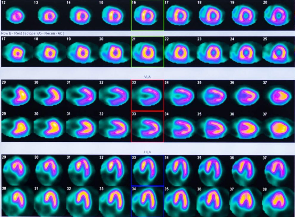
Myocardial perfusion imaging: This is a type of nuclear medicine scan that shows how well blood is flowing to the heart muscle. During the scan, a radiopharmaceutical is injected into a vein in the arm, and a special camera is used to take images of the heart as the radiopharmaceutical travels through the bloodstream. This test can help diagnose blockages or narrowing of the coronary arteries, which can cause chest pain or other symptoms.
Cardiac viability imaging: This is a type of nuclear medicine scan that helps determine if a portion of the heart muscle is still alive or has died as a result of a heart attack. During the scan, a radiopharmaceutical is injected into a vein in the arm, and images are taken of the heart to determine which areas are still functioning properly.
Cardiac sarcoidosis imaging: This is a type of nuclear medicine scan that helps diagnose sarcoidosis, an inflammatory disease that can affect the heart. During the scan, a radiopharmaceutical is injected into a vein in the arm, and images are taken of the heart to look for areas of inflammation.
Heart transplant imaging: Nuclear medicine can be used to evaluate the function of a transplanted heart. During the scan, a radiopharmaceutical is injected into a vein in the arm, and images are taken of the heart to assess its function and detect any signs of rejection.
These are just a few examples of the many ways that nuclear medicine can be used to diagnose heart diseases. Your doctor can provide more information about the specific tests and procedures that may be appropriate for your individual situation.
PET, PET-CT and D-SPECT
PET-CT, PET, and D-SPECT are different types of nuclear medicine imaging techniques that can be used to diagnose heart diseases. Overall, each of these imaging techniques has unique strengths and limitations. PET-CT combines the benefits of both PET and CT scans, providing detailed anatomical and functional information. PET is a versatile imaging technique that can be used to diagnose a wide range of heart conditions, while D-SPECT provides high-quality images with improved detection sensitivity. Your doctor can help determine which imaging technique is most appropriate for your individual situation. Here are some of the characteristics of each technique:
Positron Emission Tomography (PET)
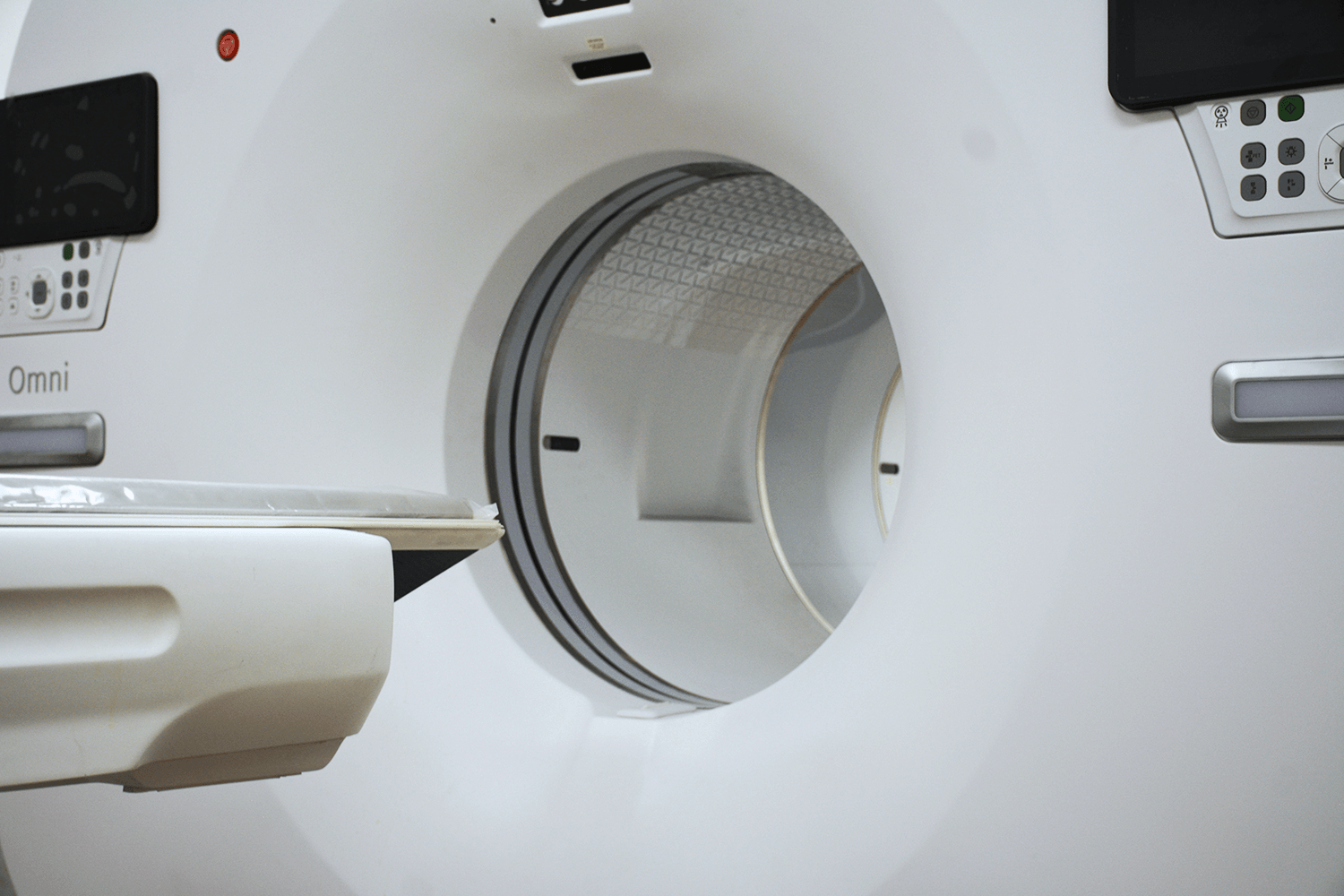
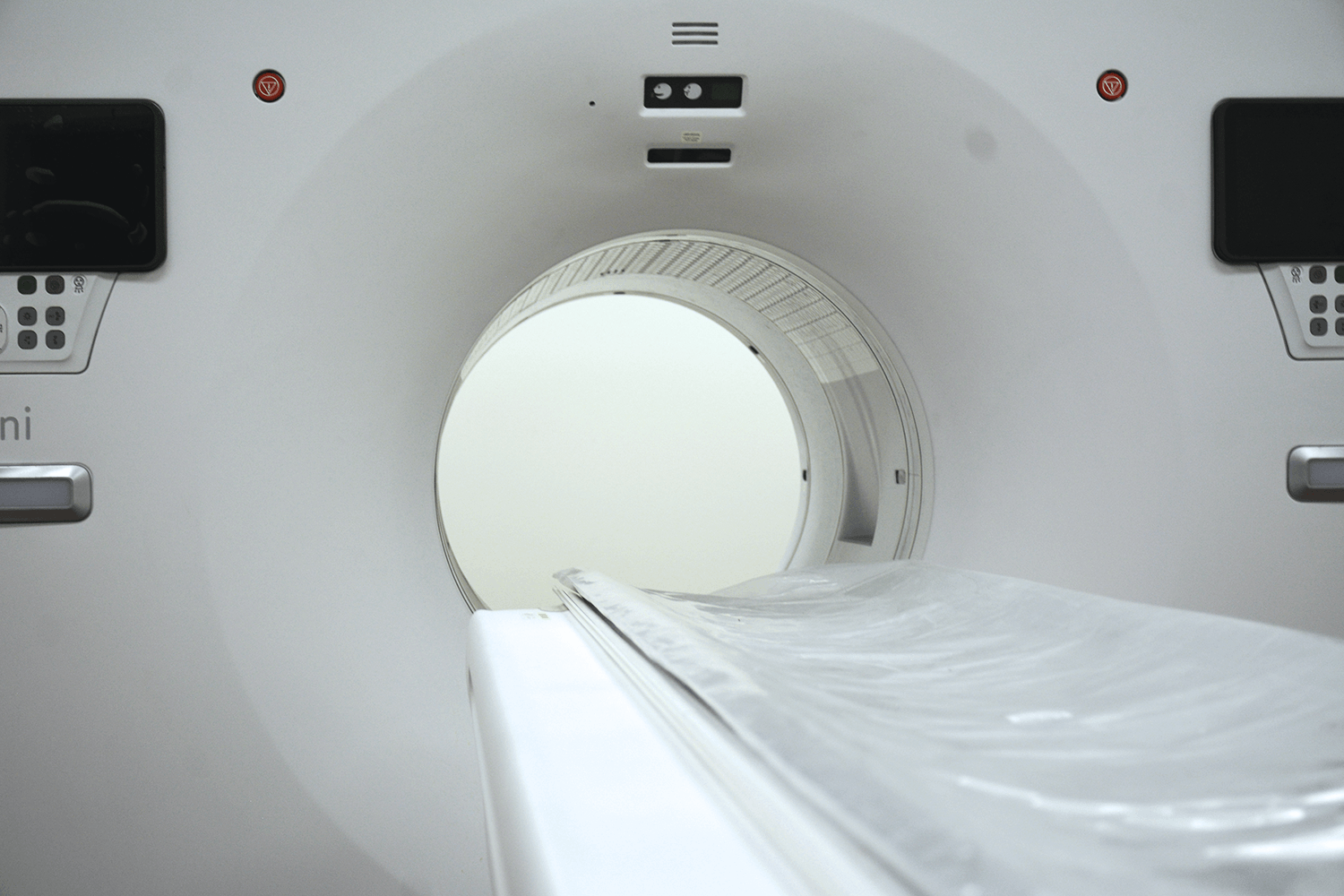
PET: Positron Emission Tomography (PET) is a nuclear medicine imaging technique that uses a radiopharmaceutical that emits positrons to create images of the heart’s metabolic activity. PET can be used to evaluate blood flow, metabolism, and oxygen consumption in the heart, and it is commonly used to diagnose heart disease, such as coronary artery disease and heart failure.
One of the main advantages of PET imaging is its ability to detect diseases in their early stages, before they cause significant damage to the body. PET can also help to determine the effectiveness of treatment by monitoring changes in the metabolic activity of tumors or other tissues.
PET imaging is generally considered safe, although it does involve exposure to a small amount of radiation from the radiotracer. The risks associated with radiation exposure are generally very low, and the benefits of the diagnostic information provided by PET often outweigh any potential risks.
Overall, PET is a valuable tool in the diagnosis and treatment of a wide range of medical conditions, providing detailed information about the metabolic activity of the body’s tissues and helping doctors to make more informed decisions about patient care.
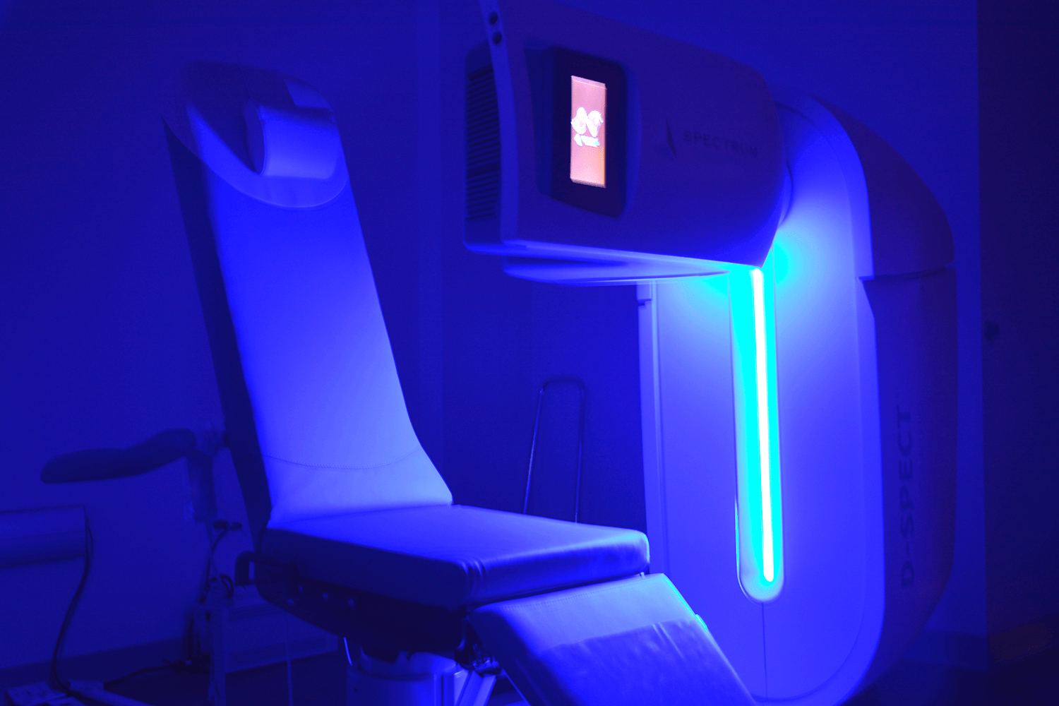
D-SPECT
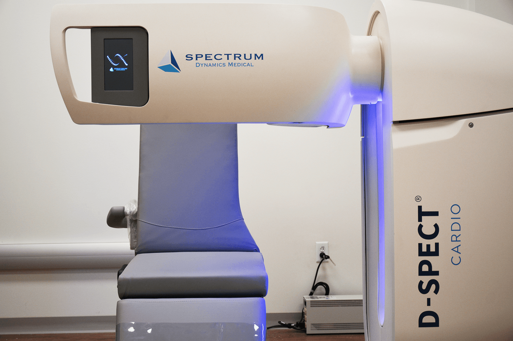
D-SPECT is a type of nuclear medicine imaging technique that uses a specialized camera to produce high-quality images of the heart. It uses a small amount of radiopharmaceutical and a dedicated detector system to produce images with high spatial resolution and improved detection sensitivity. D-SPECT can be used to diagnose various heart conditions, including myocardial ischemia, heart failure, and cardiac sarcoidosis.
Compared to traditional gamma cameras used in nuclear medicine, D-SPECT has several advantages.
Detection Sensibility: It provides higher detection sensitivity, which allows for a smaller amount of radiopharmaceutical to be used, resulting in reduced radiation exposure for the patient.
Image quality: Improved image quality, with higher spatial resolution and less image distortion.
Wide range uses: Used to diagnose various heart conditions, such as myocardial ischemia, heart failure, and cardiac sarcoidosis.
Small Radiopharma: During the test, a small amount of radiopharmaceutical is injected into a vein in the arm, and images are taken of the heart using the D-SPECT camera.
Further vision: The images can show how well blood is flowing through the heart, the size and shape of the heart chambers, and the presence of any areas of inflammation or damage.
Overall, D-SPECT is a valuable tool in the diagnosis and treatment of heart disease, providing high-quality images with improved detection sensitivity and reduced radiation exposure for the patient. However, it is important to note that D-SPECT may not be appropriate for all patients, and your doctor can help determine if it is the best imaging technique for your individual situation.
AHI works with the latest and most advanced D-SPECT technology of the market, providing the best nuclear medicine service to its patients, with Spectrum medical Dynamics as the supplier.
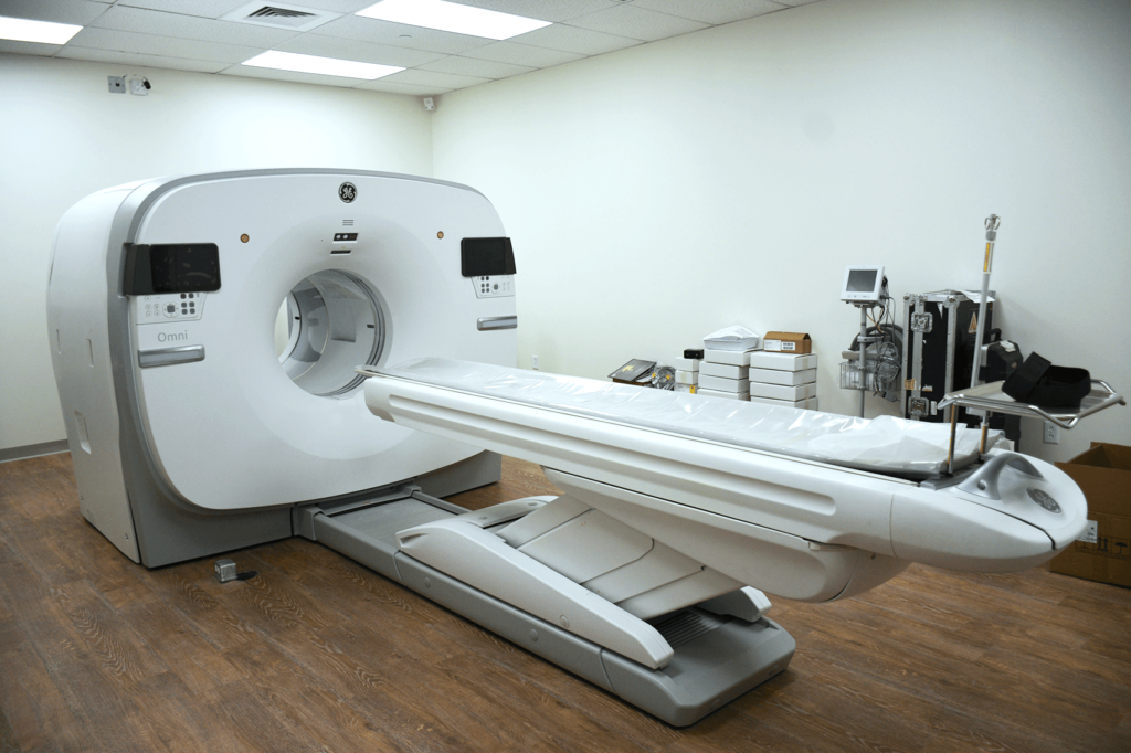
PET-CT: Positron Emission Tomography-Computed Tomography (PET-CT) is a combination of two imaging techniques that can provide detailed information about both the structure and function of the heart. PET-CT uses a radiopharmaceutical that emits positrons, which are detected by a special camera to create images of the heart’s metabolic activity. A CT scan is also performed at the same time to provide detailed anatomical information. This technique can be used to diagnose a range of heart conditions, including coronary artery disease, heart failure, and myocardial viability. Here are some of their benefits:
Accurate Diagnosis: Cardiac PET CT can provide highly accurate and detailed information about the hearts internal structures and functions, making it an effective tool for diagnosing a wide range of cardiac conditions such as coronary artery disease, heart valve disorders, and heart failure.
Early Detection: Cardiac PET CT can detect cardiac abnormalities at early stages before they become symptomatic, allowing for early intervention and treatment.
Non-invasive: Cardiac PET CT is a non-invasive imaging technique, which means it does not require any surgical incisions or invasive procedures. This reduces the risk of complications and reduces recovery time.
Comprehensive Imaging: Cardiac PET CT combines two imaging modalities, PET and CT, to produce highly detailed images that provide both functional and anatomical information about the heart and surrounding structures. This makes it easier to pinpoint the location of abnormalities and assess their severity.
Treatment Planning: Cardiac PET CT can provide valuable information to help guide treatment planning for a wide range of cardiac conditions. This includes determining the extent and severity of a disease, assessing the effectiveness of treatment, and monitoring disease progression.
Reduced Radiation Exposure: Cardiac PET CT uses a lower dose of radiation compared to traditional nuclear medicine scans, reducing the risk of radiation exposure for patients.
Overall, cardiac PET CT is a highly valuable imaging modality for the diagnosis and management of cardiac conditions. It provides accurate and detailed information about the hearts internal structures and functions, allowing for early detection and intervention. Additionally, it is non-invasive and uses a lower dose of radiation compared to traditional nuclear medicine scans, making it a safer and more convenient option for patients.
Amarillo Heart Institute works with the latest and most advanced PET-CT technology, providing the best nuclear medicine service to its patients, with GE as the supplier.


")


