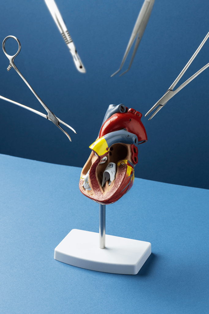
The heart never takes a break. The heart is a strong muscle that never stops exercising, not for a minute. Every minute it needs blood, nourishment and oxygen. At Amarillo Heart Institute, we understand the importance of keeping your heart healthy and functioning at its best. We believe that a healthy heart is the foundation of a healthy life, and our goal is to provide you with all you need to achieve optimal heart health, a happier heart.
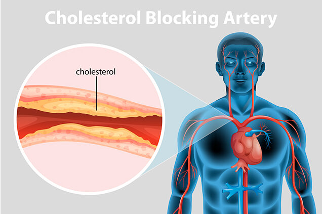
Percutaneous coronary revascularization (PCR), also known as percutaneous coronary intervention (PCI), is a minimally invasive procedure used to treat blockages in the coronary arteries, which supply blood to the heart muscle. The procedure involves the use of a catheter inserted through a small incision in the groin or wrist and threaded through the blood vessels to the heart.
During the procedure, a small balloon at the tip of the catheter is inflated to compress the plaque or blockage in the coronary artery, allowing blood to flow more freely to the heart muscle. In some cases, a stent may be placed to help keep the artery open after the balloon is deflated.
PCR is often used to treat patients with coronary artery disease (CAD), which is caused by a buildup of plaque in the arteries that supply blood to the heart. The procedure can help improve blood flow to the heart and relieve symptoms such as chest pain or shortness of breath.
PCR is typically performed in a cardiac catheterization laboratory (Cath Lab) and is usually done under local anesthesia. The procedure is generally considered safe and effective, with a low risk of complications such as bleeding or infection. However, as with any medical procedure, there are risks involved, and patients should discuss the potential benefits and risks with their healthcare provider before undergoing PCR.
Overall, percutaneous coronary revascularization is an important treatment option for patients with CAD, and has helped to improve outcomes and quality of life for many individuals with this condition.
Percutaneous coronary revascularization (PCR), also known as percutaneous coronary intervention (PCI), is a minimally invasive procedure used to treat blockages in the coronary arteries, which supply blood to the heart muscle. The procedure involves the use of a catheter inserted through a small incision in the groin or wrist and threaded through the blood vessels to the heart.
PCR is often used to treat patients with Coronary artery disease (CAD), which is caused by a buildup of plaque in the arteries that supply blood to the heart. The procedure can help improve blood flow to the heart and relieve symptoms such as chest pain or shortness of breath.
PCR is typically performed in a cardiac catheterization laboratory (Cath Lab) and is usually done under local anesthesia. The procedure is generally considered safe and effective, with a low risk of complications such as bleeding or infection. However, as with any medical procedure, there are risks involved, and patients should discuss the potential benefits and risks with their healthcare provider before undergoing PCR.
During the procedure, a small balloon at the tip of the catheter is inflated to compress the plaque or blockage in the coronary artery, allowing blood to flow more freely to the heart muscle. In some cases, a stent may be placed to help keep the artery open after the balloon is deflated.
Overall, percutaneous coronary revascularization is an important treatment option for patients with CAD, and has helped to improve outcomes and quality of life for many individuals with this condition.
Types of Percutanous Cornary Revascularization
There are several types of percutaneous coronary revascularization procedures (PCR) that can be performed depending on the severity and location of the blockage in the coronary arteries. Some of the most common types of PCR procedures include:
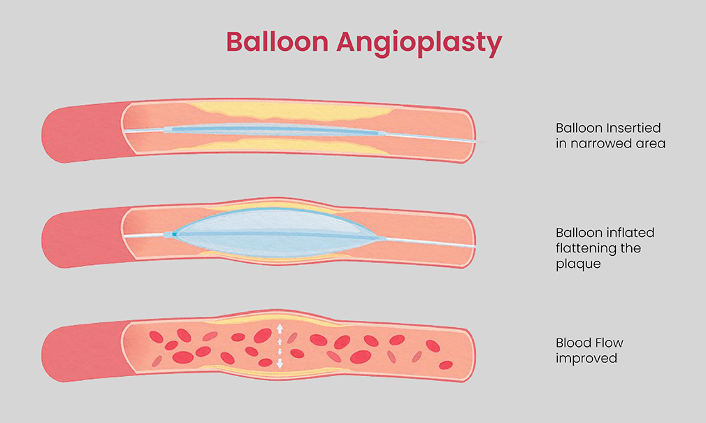
Percutaneous transluminal coronary angioplasty (PTCA) or Balloon Angioplasty
Percutaneous transluminal Coronary angioplasty (PTCA) is another name for balloon angioplasty, used to treat Coronary artery disease (CAD).
During PTCA, a catheter with a small balloon at the end is inserted into the blocked artery. The balloon is then inflated to widen the artery and compress the plaque against the artery wall. The balloon can be plain or drug-coated. Drug-coated balloons release medication to prevent the artery from narrowing again. This helps to restore blood flow to the affected area, improve symptoms such as pain or cramping, and prevent further damage to the tissue or limb.
PTCA is often performed in combination with stent placement to help keep the artery open after the balloon is deflated and removed. The procedure is typically performed under local anesthesia and takes about one to two hours to complete. Patients can usually return home the same day or the following day and resume normal activities within a week.
PTCA is a minimally invasive alternative to traditional open surgery and has a lower risk of complications and shorter recovery time. It is an effective treatment option for patients with peripheral arterial disease who are not candidates for surgery or who prefer a less invasive approach.
There are different types of balloon angioplasty, including:
High-pressure balloon angioplasty: This type of balloon angioplasty uses a higher pressure to inflate the balloon and widen the artery.
Cutting balloon angioplasty: This involves a balloon with small blades on its surface that are designed to cut the plaque during inflation, making it easier to compress against the artery wall.
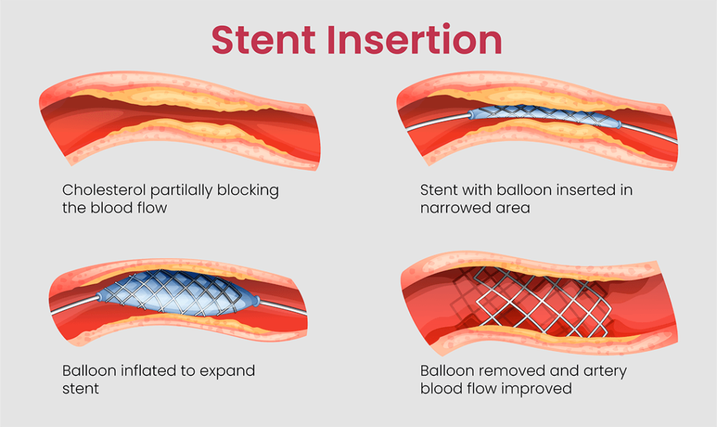
Stent insertion is a type of percutaneous coronary revascularization procedure (PCR) that is used to treat blockages in the coronary arteries. A stent is a small, mesh-like metal or plastic tube that is inserted into the artery to help keep it open and prevent it from becoming blocked again.
There are three main types of stents that can be used in stent insertion procedures: bare metal stents (BMS), drug-eluting stents (DES), and bioabsorbable stents (BAS).
Stent types:
Bare metal stents (BMS): These stents are made of a base metal such as stainless steel or cobalt-chromium. They are designed to provide structural support to the artery and prevent it from collapsing or becoming blocked again. BMS are usually less expensive than other types of stents, but they may have a higher risk of restenosis (re-narrowing of the artery) compared to drug-eluting stents.
Drug-eluting stents (DES): These stents are coated with medication that is slowly released over time to help prevent restenosis. The medication helps to inhibit the growth of scar tissue inside the artery and reduce the risk of the stent becoming blocked again. DES are typically made of a base metal such as stainless steel or cobalt-chromium, and may also be coated with a polymer material to help control the release of the medication.
Bioabsorbable stents (BAS): These stents are made of a biodegradable material such as polylactic acid (PLA) or polyglycolic acid (PGA) that gradually dissolves over time. As the stent dissolves, it is replaced by natural tissue growth, which helps to restore the normal function of the artery. BAS may have a lower risk of restenosis compared to other types of stents, but they are still relatively new and their long-term safety and effectiveness are still being studied.
During the stent insertion procedure, a catheter is inserted into the artery through a small incision in the groin or wrist and threaded up to the blocked artery. Once the stent is in place, it is expanded using a balloon catheter to compress the plaque and widen the artery. The balloon is then deflated and removed, leaving the stent in place to help keep the artery open.
Atherectomy is a minimally invasive procedure used to treat blockages in the arteries. The procedure involves removing or reducing the buildup of plaque that has formed inside the artery walls, which can impede blood flow and lead to a variety of health complications, including heart attacks and strokes.
There are four main types of atherectomy procedures, including: Directional, Rotational, Orbital and Laser.
Overall, atherectomy procedures can be highly effective in treating blockages in the arteries, and can help improve blood flow and reduce the risk of complications such as heart attacks and strokes. However, like any medical procedure, atherectomy comes with some risks, including bleeding, infection, and damage to the artery wall. It is important to talk to your doctor about the risks and benefits of atherectomy, and to discuss any concerns you may have before undergoing the procedure.
Atherectomy TYPES
Directional atherectomy: This procedure involves using a catheter with a rotating blade to remove the plaque from the inside of the artery. The blade is designed to shave off the plaque, which is then collected and removed from the body. This technique is particularly useful in removing harder, calcified plaques.
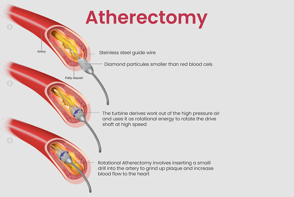
Rotational atherectomy: This procedure involves using a specialized catheter with a diamond-coated, high-speed rotating burr to remove the plaque from the inside of the artery. The burr breaks up the plaque into small particles, which are then removed from the body by the bloodstream or with the help of a suction catheter.
Orbital atherectomy: This procedure uses a diamond-coated, eccentrically mounted, high-speed rotational crown to abrade the plaque from the inside of the artery. The crown’s unique design allows it to move in an elliptical motion, reducing the risk of damaging the artery wall while still effectively removing the plaque.
Laser atherectomy: This procedure involves using a laser to vaporize the plaque inside the artery. The laser is delivered through a catheter, and the heat generated by the laser vaporizes the plaque, which is then removed from the body by the bloodstream.
Thrombectomy is a procedure used in percutaneous Coronary revascularization (PCR) to remove a blood clot (thrombus) from an artery that has become blocked. This procedure is typically performed using a catheter inserted through a small incision in the groin or arm, and is done under local anesthesia.
The catheter is guided to the site of the clot using imaging techniques such as ultrasound or fluoroscopy. Once the catheter is in place, a device is used to break up the clot into smaller pieces, which are then suctioned out of the artery. This process is called mechanical thrombectomy and is often performed using a device called a thrombectomy catheter.
Thrombectomy is typically used when the blood clot is too large or too difficult to remove using medication alone, such as with anticoagulant or thrombolytic drugs. It is also used in cases where the clot has caused tissue damage or limb-threatening ischemia.
Like other PCR procedures, thrombectomy carries some risks, including bleeding, infection, and damage to the artery or surrounding tissue. The procedure is usually performed by a vascular surgeon or interventional radiologist who has experience in performing PCR procedures.
Overall, thrombectomy is an effective treatment option for patients with peripheral arterial disease who have a blood clot that cannot be treated with medication alone. The procedure can help restore blood flow to the affected area, relieve symptoms, and prevent further damage to the tissue or limb.